Cytopathology
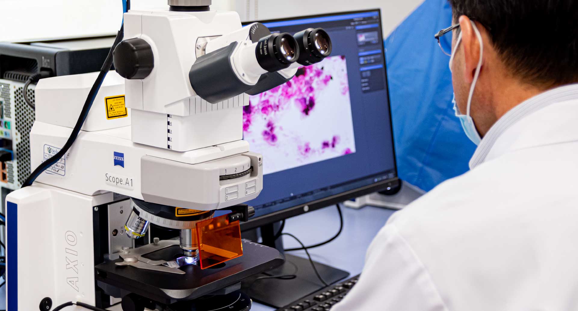
In comparison to histopathology techniques, diagnostic cytology provides a quick and relatively non-invasive way to evaluate the cellular reaction to infection, inflammation and neoplasms.
Important factors affecting the cytological picture are: the type of injury or the type of invading pathogen, the site of infection process, its route of entry and the stage of the disease i.e. acute and chronic recurrent phase. The types of cells such as epithelial cells, neutrophils, lymphocytes, monocytes, bacteria or fungi are evaluated.
The role of specific cells such as in ensuing immune response, phagocytic activity of monocytes and their activated forms in removing cell debris and pathogenic organisms, the extent of tissue damage, the stage of disease and the prognosis are assessed. Currently, Diff Quick or Giemsa stain, Gram’s stain, Papanicolaou and PAS stains are routinely used for diagnostic cytology.